High resolution imaging (HD/HDRI) and video recording (RGB/BW)
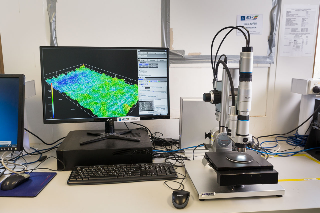
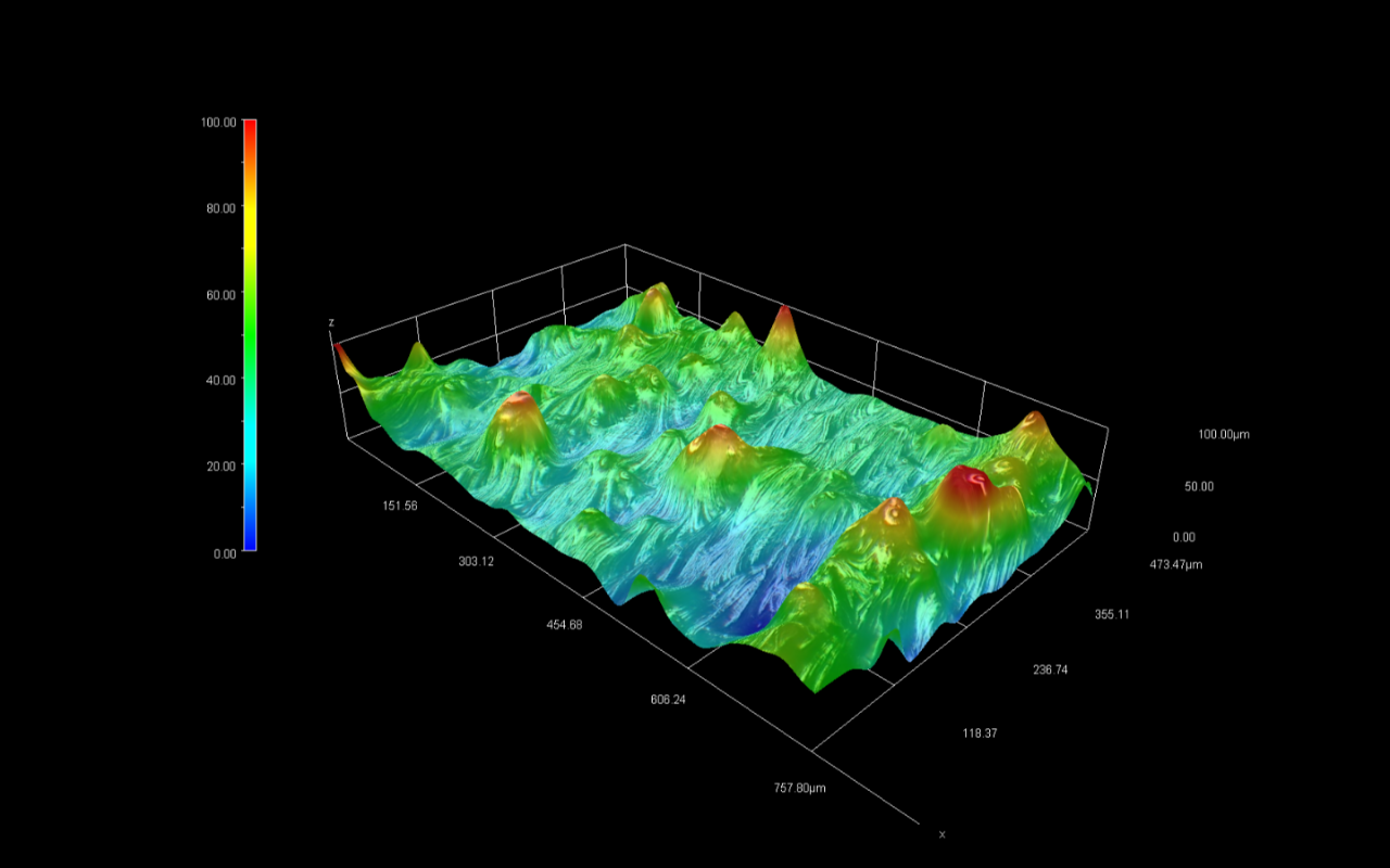
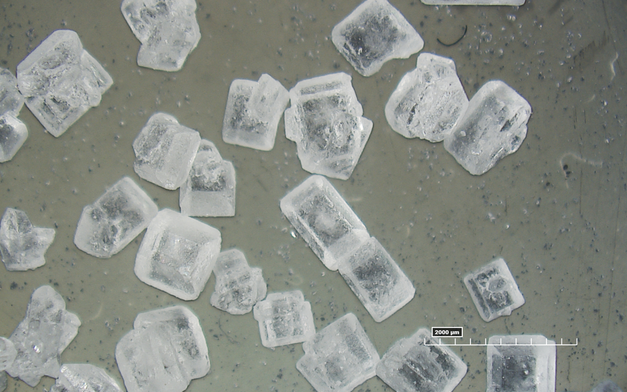
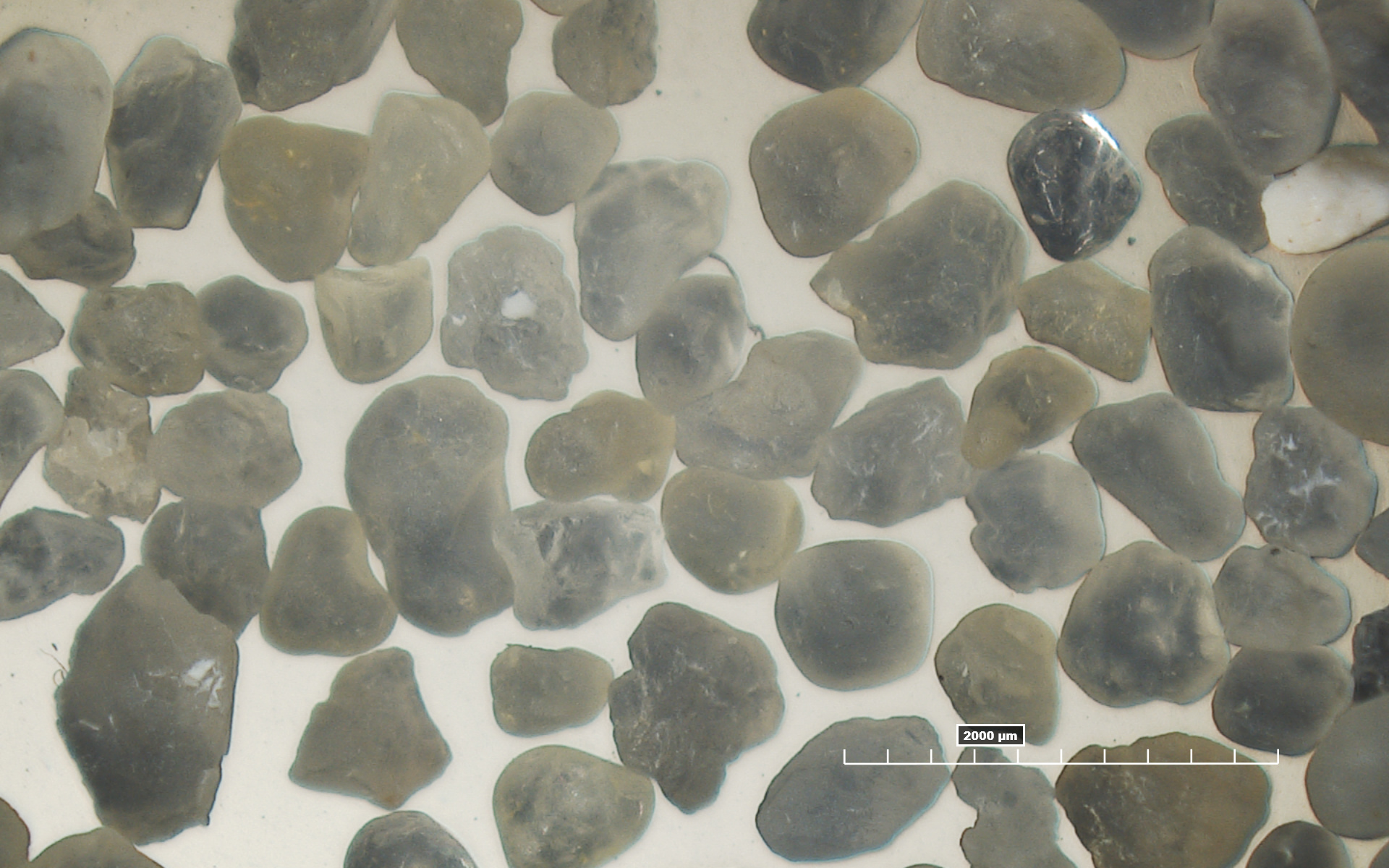
The Hirox RH-2000 2D/3D digital microscope can facilitate acquisition of high-resolution images of your samples, with calibrated 2D scales and high-quality 3D reconstructed maps, allowing cross-section profiling, roughness analysis and XYZ data export for processing and 3D printing.
Specifications
- 3D images up to 10,000×
- High intensity LED source provides true colour reproduction
- 1920×1200 pixel resolution at 50 FPS
- Motorised stage with 40mm × 40mm working range
- Up to 180° inclination on camera stage to target specific features on samples
Access
The Hirox can be operated by researchers or on a fee-for-service basis by MCFP platform staff. Trained users can book through the MCFP iLab booking system. Please contact Tian below to arrange access or training.
Additional Resources

Tian Zheng
Academic Specialist - Nanomaterials Characterisation
Tian leads the nanomaterials characterisation (NMC) node of the MCFP. She can provide insight into all modes of modern atomic force microscopy measurements on a range of samples from nanomaterials to biological structures.

See Also
-
Cypher ES AFM
High resolution (sub-nanometre) imaging, viscoelasticity mapping, electrical properties
-
Scanning electron microscopy (SEM)
The FlexSEM 1000 is a rapid and easy to use variable pressure SEM amenable to imaging both conductive and insulating specimens
-
Confocal Raman Microspectroscopy
The Renishaw inVia Qontor confocal Raman microscope is capable of spectroscopy, mapping and live focus/topography tracking