Identification of T:B Cell Multimers After Bispecific T Cell Engagement, Using Both Conventional and Imaging Flow Cytometry
Joanne E. Davis, Mandy Ludford-Menting, Helena Peng, Darryl N. Johnson, David S. Ritchie, and Rachel Koldej
Cytometry Part A (2026) https://doi.org/10.1002/cyto.a.70039
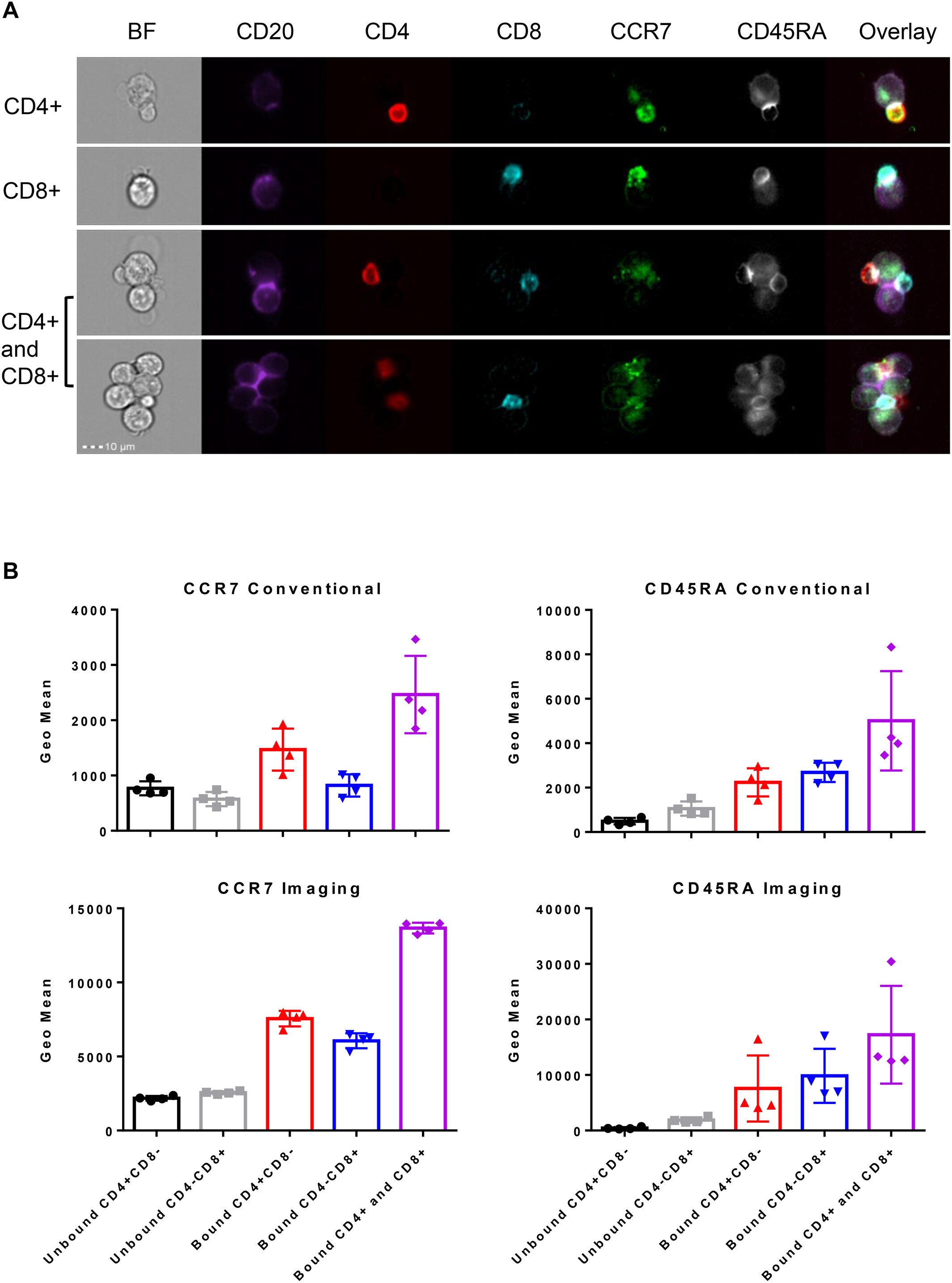
Bispecific T cell engagers bring together T cells with malignant B cell targets to enable synapse formation and disease eradication of hematological malignancies. The proportion of T cells and their ability to bind to their targets varies between patients and may be a biomarker of response. In this study, we developed a method to detect T and B cell conjugates, as measured by conventional flow cytometry, and confirmed using imaging flow cytometry. After bispecific antibody engagement, multiple T cells were bound to each target cell in complexes comprising CD4+, CD8+, and CD4+ and CD8+ T cells. Imaging flow cytometry confirmed that the number and size of multimers increased significantly in the presence of bispecific antibodies. Conventional cytometry misidentified the memory phenotype of T cells bound to target cells due to augmented co-expression of CD45RA and CCR7 cell surface memory markers. Imaging flow cytometry verified conventional flow cytometry data showing increased T:B synapse and multimer formation after incubation with bispecific T cell engagers. Therefore, both flow cytometry platforms are suitable for identification of T cell: target cell conjugates.
Our expertise and capabilities in this research
-
Darryl Johnson
Darryl leads the Cytometry node of the MCFP with expertise in mass and flow cytometry, and confocal microscopy. He has a background in molecular biology, biochemistry and immunology, and can help you with experimental design, preparation, acquisition, and interpretation.
-
Imaging Flow Cytometry
The Amnis Imagestream X MkII Imaging Flow Cytometer (ISX) bridges the gap between microscopy and flow cytometry by combining the structural information from microscopy and the high throughput nature of flow cytometry