Introduction
What is an atomic force microscope?
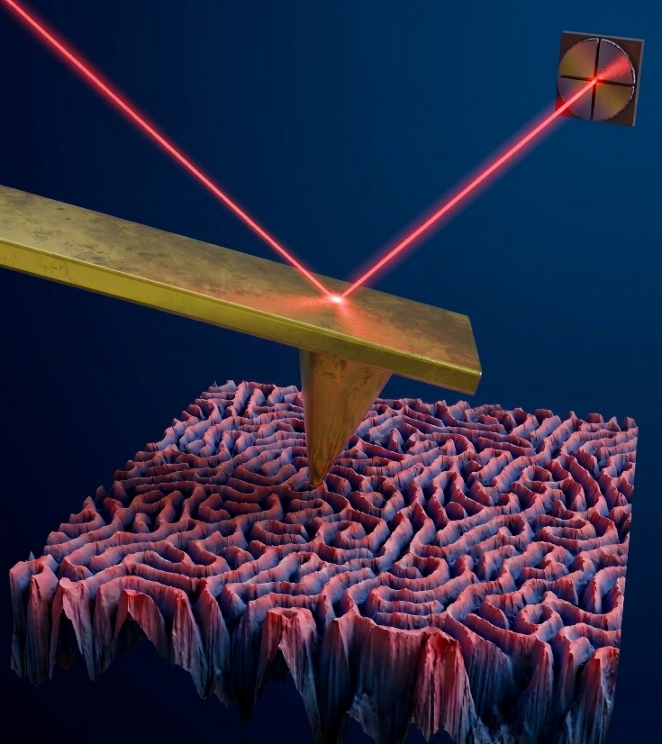
An atomic force microscope, or AFM, uses in its most basic configuration an optical beam deflection system to convert into height data the deflection of a sharp probe scanning the sample (in continuous or intermittent contact). This data can be later used to create a 3D reconstructed sample profile.
The probe, a cantilever beam with a sharp pyramid at one end, touches the sample and moves up and down in sync with the topography of the sample. Depending on the scanning mode, several data channels can be extracted: amplitude, height, and phase shift. Data in-line and post-processing can expand the range of channels to stiffness, Young’s modulus, adhesion, loss-tangent, surface potential, indentation, electrical conductivity.
Given the very low noise floor (several tens of picometres) and precise XY piezo crystals’ motion, one can scan, large areas, such as 100×100 µm2, or very small domains, 5×5 nm2. Such versatility allows detecting atomic lattices and Moiré patterns in air and in fluids. Z resolution reaches tens of pm and typical systems can handle samples with peak-to-valley dimensions up to 12 µm.
These are your AFMs
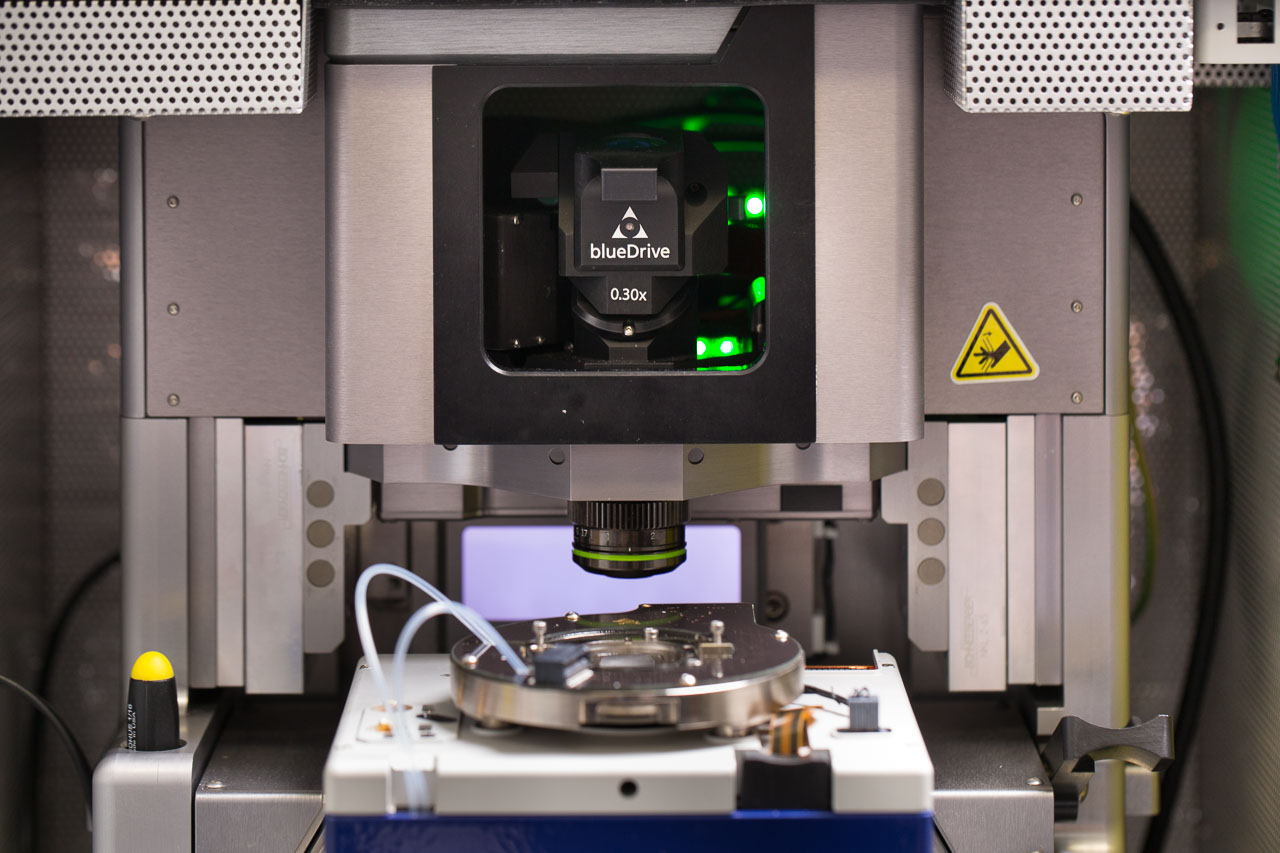
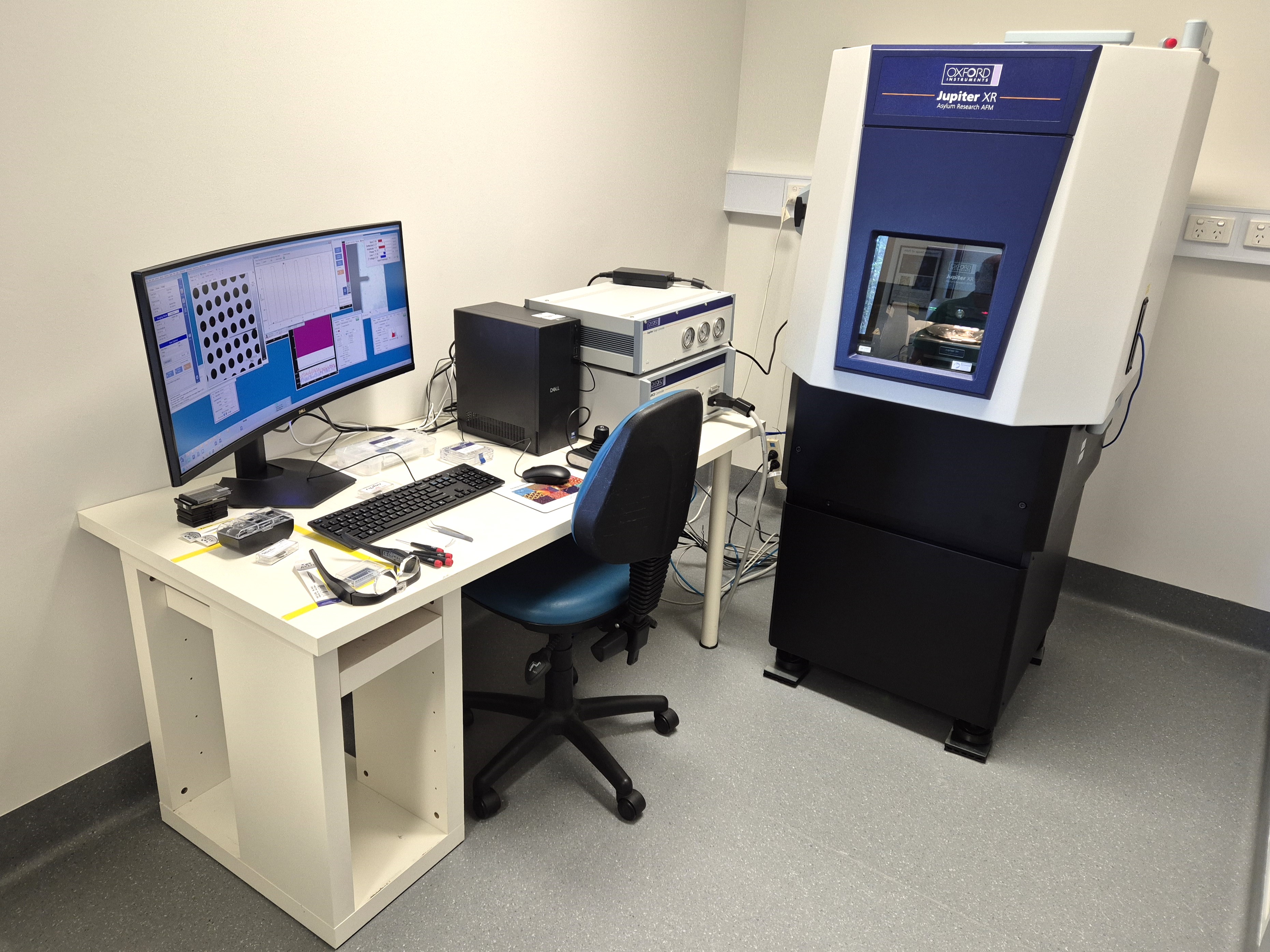
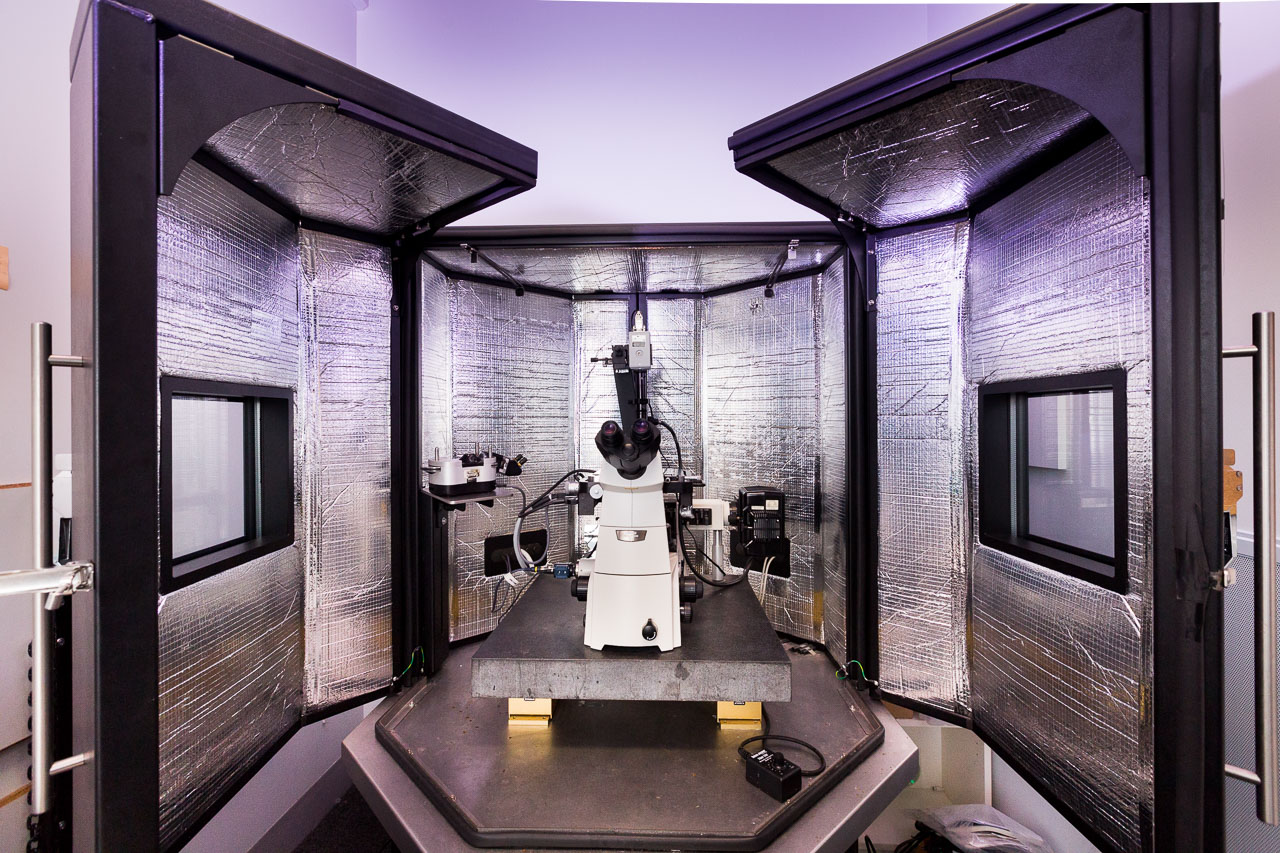
The MCFP operates three Asylum Research/Oxford Instruments AFMs
Capabilities
While each of our instrument excel at AFM, there are specific strengths in their capabilities.
-
Probe actuation Holders Gas Fluid Temperature XY Scan Size Z Scan Range
Piezo
BlueDrive (640 nm)Air
Dual ORCAAir Droplet No control 100×100 μm
Stitching±6 μm -
Probe actuation Holders Gas Fluid Temperature XY Scan Size Z Scan Range Piezo
BlueDrive (405 nm)Air/gas/fluid perfusion
ORCAAir
NitrogenDroplet
PerfusionHeating to ≤120 °C 30×30 μm
±1.5 μm -
Probe actuation Holders Gas Fluid Temperature XY Scan Size Z Scan Range Piezo Air/fluid
Air Droplet
PerfusionBioheater 90×90 μm
±5.0 μm
Throughout this module you will learn more about the fundamentals of AFM, and how these capabilities are applied in scientific research.